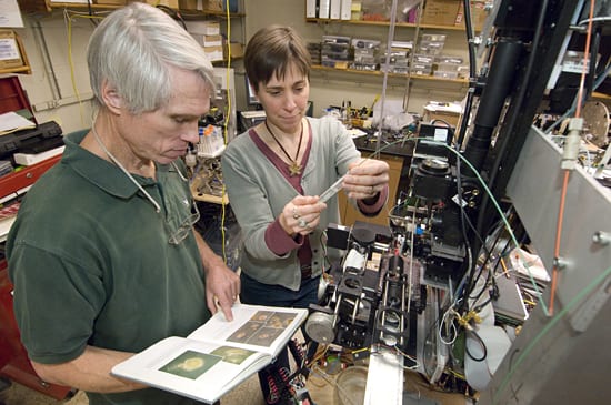
WHOI biologists Rob Olson and Heidi Sosik examine plankton-filled water samples on a prototype version of the Imaging FlowCytobot (IFCB) in Olson’s Woods Hole laboratory. The Cytobot, which is automated and submersible, counts microscopic plants in the water and photographs them. The images and data are relayed by cable to a shore-based laboratory, where specially developed software classifies the plankton into taxonomic groups. The instrument was recently used to detect a bloom of harmful marine algae (Dinophysis acuminata) in the Gulf of Mexico and prevent human consumption of tainted shellfish. (Photo by Tom Kleindinst, Woods Hole Oceanographic Institution)
More Content From :
Ocean Tech


Image and Visual Licensing
WHOI copyright digital assets (stills and video) on this website can be licensed for non-commercial use upon request and approval. Please submit your request via our Media Request Form.
For assistance or accessibility accommodations, call (508) 289-2647.